The human ear is the highly advanced result of millions of years of evolutionary progress. Everyone knows that the ear is the organ used for hearing, but not many people are aware that it’s also necessary for balance.
Most people also consider the ear just one part of the human body, but it’s a complex organ composed of many smaller, finely-tuned parts. This article describes the anatomy of the ear in-depth and discusses ways to discover and correct potential hearing disorders.
What Is the Anatomy of an Ear?
The ear is an unusually complex organ in human anatomy. Don’t worry, though—each part has a purpose that is easy to understand. In this section, we describe the anatomy of the ear in simple terms.
External Ear Anatomy (Auricle or Pinna)
The outer ear auricle or external ear is composed of all of the parts of the ear outside the skull. It is also sometimes referred to as the auricle or the pinna.
Although the outer ear is the least important part of the ear’s hearing function, it provides the necessary structure and protection. Its dish-like shape is also essential for collecting sound waves. This sound collection is the primary purpose of all of the parts of the external ear auricle anatomy.
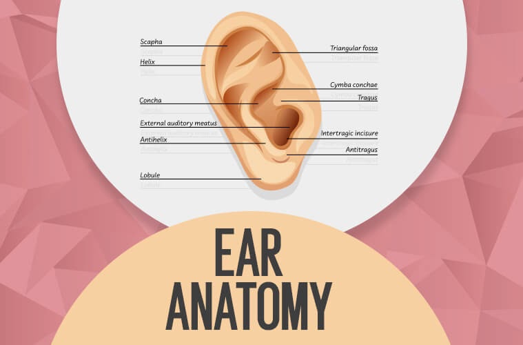
Helix
The outer, external ear can be loosely described as two cartilaginous loops, a small one nestled inside a larger one. The superior crus is the larger of the two loops, and it’s the very flexible outer curve of the ear.
Antihelix
The antihelix is the smaller of the two loops. It is more rigid and provides more strength to the outer ear. It looks somewhat Y-shaped, and the two forks at the top of the ‘Y’ are called the superior crus and the inferior crus.
Superior Crus
The superior crus is the larger, upper branch of the ‘Y’ part of the antihelix. It is shallow and generally not well-defined.
Inferior Crus
The smaller, lower branch of the antihelix ‘Y’ is called the inferior crus. It is more sharply defined, and it partially marks the border of the concha.
Concha
The concha is the smaller cavity that funnels sound waves into the ear canal. It is bordered by the antihelix, the tragus, and the antitragus. Hearing amplification devices, such as MD HearingAids or other services, are typically nestled within this cavity.
Obstruction in the concha can lead to hearing problems or conditions.
External Auditory Meatus
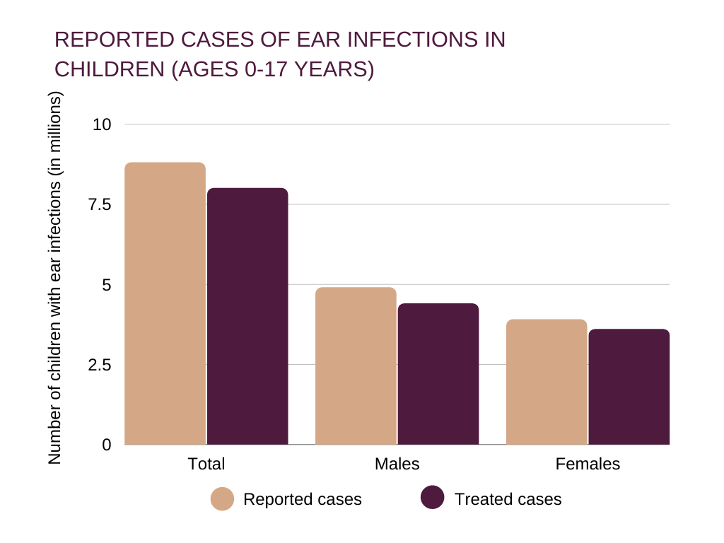
The external auditory meatus, or ear canal, is a narrow canal that leads from the concha to the tympanic membrane, or eardrum. Sound waves are delivered through this canal. This canal is prone to ear infections.
Tragus
This is the small, rigid part of the ears along the front of the ear, adjacent to the face. It borders the concha and partially shields it from debris and particularly loud noises.
Antitragus
The antitragus is the cartilage that forms the bottom part of the ‘Y’ of the antihelix. It provides a shelf-like platform at the base edge of the concha that can support the weight of medical devices. Check out our best hearing aids review on this site.
Lobule
The lobule, or ear lobe, is the fleshy skin in position below the antitragus. Experts have not reached a consensus on the purpose of this part of the ear. However, some believe its large blood supply can aid in keeping the ear warm.
Tympanic Membrane or Eardrum
The tympanic membrane, or eardrum is the final hearing organ in the outer ear, separating it from the middle ear. The eardrum collects sound waves and vibrates, passing the sound waves into the middle ear.
Most hearing disabilities are caused by trauma or disorders in the tympanic membrane eardrum. If you suspect that you are experiencing hearing problems due to tympanic membrane eardrum damage, we have listed some of the best online hearing tests that you can take to self-diagnose your hearing loss.
Middle Ear Anatomy
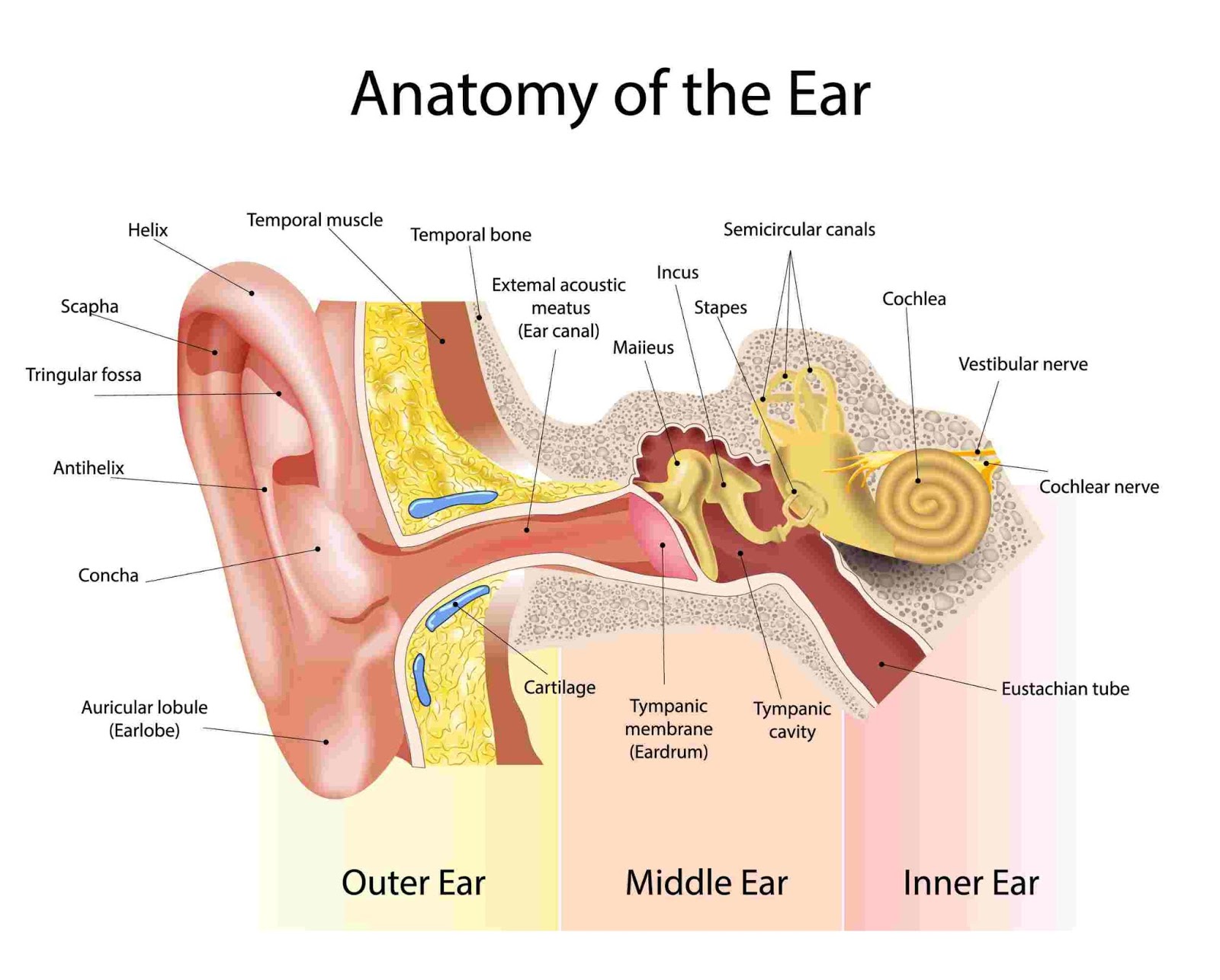
The middle ear contains most of the small organs responsible for collecting and clarifying external sound waves. It also maintains air pressure balance in the skull through the regulation of the Eustachian tube.
The middle ear is most susceptible to ear infections. It can also develop otitis media, which is a category of ear inflammation.
Tympanic Cavity
The tympanic cavity is the small space in the middle ear between the tympanic membrane ear drum and the inner ear hearing organ. This space holds the ossicles and the Eustachian Tube.
Ear infections can occur in the tympanic cavity.
Ossicles
Ear ossicles, by definition, are three very delicate bones in the middle ear that are connected like a chain. These bones oscillate in a coordinated manner to transmit sound waves and vibrations through the middle ear.
- The malleus is the largest of the ossicle bones, and due to its function, it is often called the hammer or mallet. One end of the malleus is attached to the tympanic membrane, which is functionally similar to a drum membrane. The other end is attached to the incus.
- The incus is often called the anvil. This tiny bone in the middle ear is attached to the tympanic cavity’s inner wall, and it articulates based on the vibrations and oscillation of the malleus. The opposite end of the incus is attached to the stapes.
- The stapes is the tiniest bone in the human body—with the base located in the oval window, which is one of the two membranes that separate the middle ear from the inner ear.
Eustachian Tube
The Eustachian tube travels through the middle ear from the ear cavities to the nasal cavities and the upper part of the throat. Its purpose is to equalize air pressure balance in the tympanic cavity to allow proper soundwave propagation.
When the tympanic cavity has over-pressure or under-pressure, it creates a stuffy or aching discomfort and blocked hearing. This middle ear pressure imbalance can usually be alleviated by yawning or working the jaw, which exercises the mastoid bone and opens the eustachian tube.
COVID 19 can occasionally enter the middle ear through the Eustachian tube. If you have been exposed to COVID 19, seek appropriate treatment or services.
Inner Ear Anatomy
The inner ear is where the sound waves are translated into types of electrical nerve impulses. Most of the hearing and balance content is located within the bony labyrinth. After the tympanic membrane, these are the nerves that most likely contribute to hearing impairment and may require treatment or medical services [1].
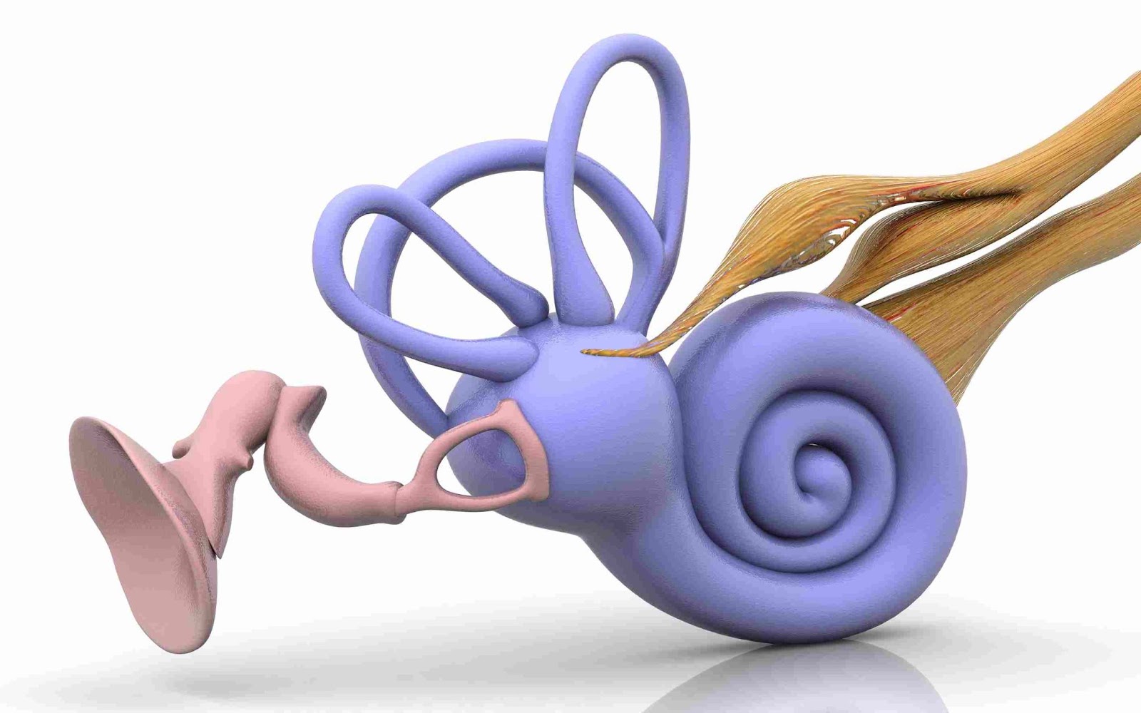
Bony Labyrinth
The bony labyrinth is located in the temporal bone on the side of the skull. It is a series of cavities that house the vestibule content, the cochlea, and the three semicircular canals.
Cell carcinoma can sometimes develop in the temporal bone and should be treated by health services.
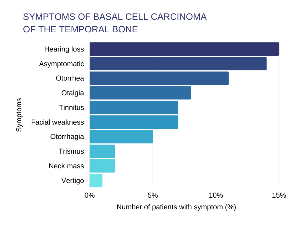
Hearing loss was the most common symptom reported among patients with basal cell carcinoma of the temporal bone
Vestibule
The vestibule is the central cavity within the inner ear. In addition to connecting the semicircular canal and the cochlea, it also contains the saccule and the utricle, two parts of the membranous labyrinth system.
Semicircular Ducts (Define Anterior, Lateral, and Postular)
The semicircular ducts, or semicircular canals, are all three looping, fluid-filled tubes necessary to maintain balance [2].
These canals form semicircular loops oriented in all three perpendicular dimensions. Tiny hair cell receptors inside each fluid-filled tube are sensitive to the fluid’s motion and momentum, and this nerve can detect very small changes in orientation to help the body maintain balance. Effectively, these nerves operate on the same principles as gyroscopic accelerometers [3].
- The lateral, or horizontal tube detects turning and twisting along the axis perpendicular to the ground (neck turning motions) and notifies the brain.
- The superior, or anterior canal tube detects back and forth rotations of the head, such as you would experience when nodding your head up and down.
- The postular duct detects side-to-side motion, when you tilt your head to the left or right, or move your body in sideways rotation (like a cartwheel).
Cochlea
The cochlea is the organ that directly translates sound vibrations into electrical nerve information.
It is a spiral-shaped cavity in the bony labyrinth, divided into three chambers.
- The scala vestibuli, or vestibular duct, is attached to the oval window.
- The scala tympani (tympanic chamber) terminates at the round window, which is the other membrane that connects to the middle ear sections.
- The scala media, or cochlear duct, contains the nerves that receive all signals from the hair cell receptors and send them to the brain.
These chambers are filled with perilymph fluid content and tiny organs called hair cell receptors. These hair cell receptors directly translate vibrations into electrical nerve information for the cochlear nerve to transmit to the brain.
FAQ
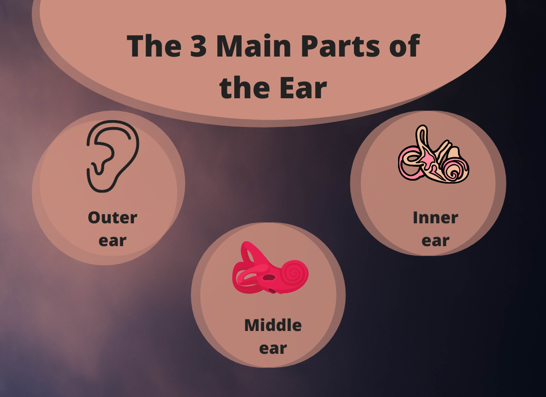
What Are the 3 Main Parts of an Ear?
In the simplest terms, the ear can be categorized into three primary anatomical collections: the outer ear, the middle ear, and the inner ear:
- The Outer Ear is the curved part of the ear that is visible on the outside of the head. Its primary purpose is to collect sound waves, just like a satellite dish collects radio waves. The outer ear is composed mostly of flexible tissue that makes it a more durable protrusion from the sides of the head.
- The Middle Ear is where most of the outside sound is picked up. Its content consists of the tympanic membrane, or eardrum, and several small, delicate bone tubes that amplify and refine sound wave vibrations.
- The Inner Ear section converts the mechanical sound waves into neural signals the brain can understand. Its content consists of sacs that transmit the sound waves to tiny hair cells. These hair cell receptors transform the movement into electrical information, much like a strain gauge, or electromechanical transducer.
What Are the 7 Main Parts of an Ear?
The ear can be further analyzed by seven content components, divided into the three ear categories listed above.
- The Pinna is the main structure of the outer ear. Composed of durable and flexible tissue, the pinna is the curved dish-like organ that gathers outside sound waves and draws them into the ear.
- The Ear Canal is the tube that connects the pinna to the inner ear. It’s only a few centimeters long, and usually less than a centimeter in diameter.
- The Tympanic Membrane is the primary organ that measures sound wave vibrations. It also divides the outer ear from the middle ear. It then transmits a clarified, amplified sound signal to the inner ear components.
- The Ossicles (malleus incus stapes) are the small, connected bones inside the middle ear that transmit sound signals from the tympanic membrane to the next area.
- The Oval Window is the membrane between the middle ear and the inner ear, specifically the cochlea.
- The Cochlea is the organ that directly translates sound signals into electrical information for the cochlear nerve to transmit to the brain. It is filled with hair cells that are extremely sensitive to vibration.
- The Semicircular Canals are a system of three fluid-filled loops. These loops are oriented in three perpendicular directions, and hair cells inside each loop detect movement and momentum, which allows you to maintain balance (similar to a gyroscopic accelerometer).
What Are the 6 Ear Bones?
Each ear contains three ossicles, or bones, in the middle ear: the malleus incus stapes. These ossicles transmit sound signals from the outer ear to the inner ear.
Conclusion
Ear anatomy might seem complex, but can be easily understood when broken down into easy-to-understand terms. It should be a helpful starting point for students hoping to understand the complex workings of the human ear.
There are also many hearing disorders that the reader should be familiar with, leading to hearing loss. We have provided helpful links to the best online hearing tests so that readers can diagnose their hearing loss. If you are suffering from a hearing disability, consider checking out MD HearingAid and their helpful products and services.
Reference
- “Hearing Loss.” Mayo Clinic, Mayo Foundation for Medical Education and Research, 10 May 2019, www.mayoclinic.org/diseases-conditions/hearing-loss/symptoms-causes/syc-20373072.
- Löwenstein O and Sand Alec 1940The mechanism of the semicircular canal. A study of the responses of single-fibre preparations to angular accelerations and to rotation at constant speedProc. R. Soc. Lond. B129256–275. http://doi.org/10.1098/rspb.1940.0039
- Zeng H, Zhao Y. Sensing Movement: Microsensors for Body Motion Measurement. Sensors. 2011; 11(1):638-660. https://doi.org/10.3390/s110100638